What is an Infection?

Physical barriers and the immune system defend the body against organisms that can cause infection. Physical barriers include the skin, mucous membranes, tears, earwax, mucus, and stomach acid.
Also, the normal flow of urine washes out microorganisms that enter the urinary tract. The immune system uses white blood cells and antibodies to identify and eliminate organisms that get through the body's physical barriers (see Biology of the Immune System).
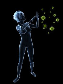
Infections are caused when pathogenic organisms invade a body. An infection is defined as the body’s response to germs that have attack the body or parts of the body. Germs that enter the body can mount an attack upon the cells, nerves and organs of the body. The response of the body when part is under attack manifests as dead or damaged cells, swelling and fever. Depending upon where the invasion is located, the symptoms of infections may be minor and in single area, or may be catastrophic and spread throughout the body.
Causes of Infection
There are four major types of infections: bacteria, viruses, fungi and protozoa. Infections caused by bacteria include sore throat, ear infections, boils and sinusitis; Viruses cause such reactions as measles, mumps, chicken pox, flu and polio. An infection caused by fungi might show up as athlete’s foot and other itchy rashes that show up in damp skin folds. Protozoa cause such diseases as malaria, thanks to the intestinal worms that invade the body. Treatment for each of the major causes for symptoms will depend largely upon the significance of the organism doing the attack and the parts of the body that are under attack.
Typical Infection Sites
Public Bathroom
Infections can be found in almost every part of the body. Although living creatures are incredibly complex and have powerful defense mechanisms built in, there are many ways in which infections can be contracted. It is amazing that people and animals are not killed or sickened due to attacks by harmful organisms. An Infection may occur any time there is a break in the skin’s surface. The skin is the largest organ and serves as the major preventative wall to the body. Cuts, scrapes and scratches are common ways by which destructive organisms can enter the body.
Physical Barriers
Usually, the skin prevents invasion by microorganisms unless it is damaged—for example, by an injury, insect bite, or burn. Other effective physical barriers are the mucous membranes, such as the linings of the mouth, nose, and eyelids. Typically, mucous membranes are coated with secretions that fight microorganisms. For example, the mucous membranes of the eyes are bathed in tears, which contain an enzyme called lysozyme that attacks bacteria and helps protect the eyes from infection.The airways filter out particles that are present in the air that is breathed in. The walls of the passages in the nose and airways are coated with mucus. Microorganisms in the air become stuck to the mucus, which is coughed up or blown out of the nose. Mucus removal is aided by the coordinated beating of tiny hairlike projections (cilia) that line the airways. The cilia sweep the mucus up the airways, away from the lungs.
The digestive tract has a series of effective barriers, including stomach acid, pancreatic enzymes, bile, and intestinal secretions. The contractions of the intestine (peristalsis) and the normal shedding of cells lining the intestine help remove harmful microorganisms.
The bladder is protected by the urethra, the tube that drains urine from the body. In males older than 6 months, the urethra is long enough that bacteria are seldom able to pass through it to reach the bladder, unless the bacteria are unintentionally placed there by catheters or surgical instruments. In females, the urethra is shorter, occasionally allowing external bacteria to pass into the bladder. The flushing effect as the bladder empties is another defense mechanism in both sexes. The vagina is protected by its normal acidic environment.
The Blood
 One way the body defends against infection is by increasing the number of certain types of white blood cells (neutrophils and monocytes), which engulf and destroy invading microorganisms. The increase can occur within several hours, largely because white blood cells are released from the bone marrow, where they are made. The number of neutrophils increases first. If an infection persists, the number of monocytes increases. The blood carries white blood cells to sites of infection. The number of eosinophils, another type of white blood cell, increases in allergic reactions and many parasitic infections, but usually not in bacterial infections.
Certain infections, such as typhoid fever, actually lead to a decrease in the white blood cell count, but how these infections cause the decrease is not known.
Phagocytosis
Phagocytosis
Phagocytosis is mediated by macrophages and polymorphonuclear leucocytes.
Phagocytosis involves the ingestion and digestion of the following:
- microorganisms
- insoluble particles
- damaged or dead host cells
- cell debris
- activated clotting factors
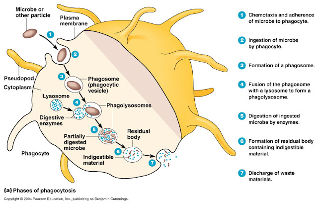
There are several stages of phagocytosis:
1. Chemotaxis
This is the movement of cells up a gradient of chemotactic factors. It may be directly induced by a substance such as C5a, produced as a result of complement activation. It can also be indirectly induced as a consequence of release of preformed mediators within mast cells by the action of C3a or C5a e.g. eosinophil chemotactic factor, or neutrophil chemotactic factor. Leukotrienes, produced by the metabolism of mast cell arachidonic acid, are also chemotactic.
2. Adherence
This works reasonably well for whole bacteria or viruses, but less so for proteins or encapsulated bacteria. In order to deal more effectively with encapsulateed bacteria, antibodies directed against the capsule enable the phagocytic cells to ingest the organisms, using their Fc receptors (see below).
3. Pseudopodium formation
This is the protrusion of membranes to flow round the "prey".
4. Phagosome formation
Fusion of the pseudopodium with a membrane enclosing the "prey" leads to the formation of a structure termed a phagosome.
5. Phago-lysosome formation
The phagosome moves deeper into the cell, and fuses with a lysosome, forming a phago-lysosome. These contain hydrogen peroxide, active oxygen species (free radicals), peroxidase, lysozyme and hydrolytic enzymes. This is known as the oxidative burst, and leads to digestion of the phagolysosomal contents, after which they are eliminated by exocytosis. Some peptides however, undergo a very important separate process at this stage. Instead of being eliminated, they attach to a host molecule called MHC class II and end up being expressed on the surface of the cell within a groove on the MHC molecule (antigen presentation).
The speed of phagocytosis can be increased markedly by bringing into action two attachment devices present on the surface of phagocytic cells:
Fc receptor: which binds the Fc portion of antibody molecules, chiefly IgG. The IgG will have attached the organism via its Fab site.
Complement receptor: the third component of complement (C3) also binds to organisms and then attaches to the complement receptor.
This coating of the organisms by molecules that speed up phagocytosis, is termed 'opsonization', and the Fc portion of antibody, and C3 are termed 'opsonins'.
The lymphatic system
"Shotty lymph nodes" refers to clusters of small swollen nodes. Shotty nodes may occur when the immune system is reacting to an infection -- it doesn't necessarily point toward any particular disease.
"Bulky disease" generally describes a lymph node or extranodal tumor that measures greater than ten centimeters in any dimension.
calcified nodes are not really rare, and typically are the result of some past, healed infection. TB is well-known to cause them, for example. Cancer is not."
Cervical lymph nodes are lymph nodes located in and around the neck.
Lymph nodes in head and neck
1: Submental
2: Submandibular
3: Supraclavicular
4: Retropharyngeal
5: Buccal
6: Superficial cervical
7: Jugular
8: Parotid
9: Retroauricular & occipital
The lymphatic system consists of organs, ducts, and nodes. It transports a watery clear fluid called lymph.
This fluid distributes immune cells and other factors throughout the body. It also interacts with the blood circulatory system to drain fluid from cells and tissues.
The lymphatic system contains immune cells called lymphocytes, which protect the body against antigens (viruses, bacteria, etc.) that invade the body. See more on lymphocytes below. It is abnormal cells of this type that cause lymphoma.
Main functions of the lymphatic system
"to collect and return interstitial fluid, including plasma protein to the blood,
and thus help maintain fluid balance,
to defend the body against disease by producing lymphocytes,
to absorb lipids from the intestine and transport them to the blood."
Role of the lymphatic system in fat absorption and transport
The lymphatic circulation as a drainage system
Lymph organs
Include the bone marrow, lymph nodes, spleen, and thymus. Precursor cells in the bone marrow produce lymphocytes. B-lymphocytes (B-cells) mature in the bone marrow. T-lymphocytes (T-cells) mature in the thymus gland.
Besides providing a home for lymphocytes (B-cells and T-cells), the ducts of the lymphatic system provide transportation for proteins, fats, and other substances in a medium called lymph.
Lymph nodes: "Human lymph nodes are bean-shaped and range in size from a few millimeters to about 1-2 cm in their normal state.
They may become enlarged due to a tumor or infection. White blood cells are located within honeycomb structures of the lymph nodes. Lymph nodes are enlarged when the body is infected due to enhanced production of some cells and division of activated T and B cells.
In some cases they may feel enlarged due to past infections; although one may be healthy, one may still feel them residually enlarged."
Lymph
"Means clear water and it is basically the fluid and protein that has been squeezed out of the blood (i.e. blood plasma). The lymph is drained from the tissue in microscopic blind-ended vessels called lymph capillaries.
These lymph capillaries are very permeable, and because they are not pressurized the lymph fluid can drain easily from the tissue into the lymph capillaries.
As with the blood network the lymph vessels form a network throughout the body, unlike the blood the lymph system is a one-way street draining lymph from the tissue and returning it to the blood."
"Unlike the cardiovascular system, the lymphatic system is not closed and has no central pump." wikipedia.org "Lymph movement occurs despite low pressure due to peristalsis - smooth muscle and skeletal activity (everyday activity and motion of the body).
"Secondary lymphatic tissues control the quality of immune responses. Differences among the various lymphatic tissues significantly affect the form of immunity and relate to how antigens (bacteria, virus, fungus, etc.) are acquired by these organs.
- Lymph nodes are filters of lymph
- the spleen is a filter of blood
- mucosal associated lymphatic tissues acquire antigens by transcytosis to lymphoid tissue from the "external" environment across specialized follicle-associated epithelial cells." Source geocities.com
"Lymphatics are found in every part of the body except the central nervous system. The major parts of the system are the bone marrow, spleen, thymus gland, lymph nodes, and the tonsils. Other organs, including the heart, lungs, intestines, liver, and skin also contain lymphatic tissue." gorhams.dk
Lymphoma
Is a disease in which malignant lymphocytes grow too fast or live too long. These cells may then accumulate in the lymph nodes or other areas of the lymphatic system to form tumors. When these cells accumulate in lymph nodes it's often called adenopathy - the enlargement of the lymph nodes; but adenopathy can have other causes.
Reactive Lymph nodes
"LYMPH nodes are a combination of burglar alarm and West Point. Like a burglar alarm they are on guard against intrusive antigen. Like West Point, the nodes are in the business of training a militant elite:" pleiad.umdnj.edu
Waxing, Waning, and Persistently Enlarged Lymph Nodes
Lymph nodes can increase or decrease in size for many reasons, including response to treatment, progression of lymphoma, spontaneous regression of lymphoma, immune activation against lymphoma, infection or the resolving of infection (reactive, see above), and so on.
Therefore, imaging of lymphoma is only an estimate of treatment response and disease direction.
Collapsing of necrotic areas in lymph nodes may explain a sudden decrease in a large lymph node.
Following treatment "a residual mass persisting on CT after treatment poses a common clinical dilemma: it may indicate the presence of viable lymphoma, which requires further treatment, or it can be benign, consisting of only fibrotic and necrotic tissues."
For this reason PET or Gallium scans may be used after treatment to help differentiate active disease from scar tissue.
Reactive lymph nodes - Lymph nodes may enlarge when immune cells react to pathogen such as virus or bacteria. Swollen glands, common to many illnesses is an example of nodes enlarging in response to a pathogen.
Lymphocytes
Lymphocytes, also called white blood cells, are described below. These are small cells, 7-9 µm in diameter in blood smears ...
These cells are the second most common white blood cell type, comprising about 30 % of the leukocyte population in peripheral blood....
Lymphocytes travel in the blood, but they routinely leave capillaries and wander through connective tissue. Therefore, lymphocytes may be normally encountered at any time in any location. They even enter epithelial tissue, crawling between the epithelial cells. They reenter circulation via lymphatic system channels (hence their name)."
Source: Blood Cells www.siumed.edu
Life cycle of lymphocytes
Develop in the thymus gland or bone marrow.
B-cells grow (differentiate) and mature in the bone marrow.
T-cells also start out in the bone marrow. They differentiate and mature in the thymus gland (beneath breastbone).
B-cell and T-cell lymphocytes are distributed through the blood stream, which eventually branches into tiny blood vessels called capillaries.
Some lymphocytes migrate to capillaries into surrounding tissues. Some enter lymphatic vessels-tiny, blind-ended tubes-and lead to larger lymphatic ducts and branches.
Along the way, the fluid passes through lymph nodes, oval structures composed of lymph vessels, connective tissue, and white blood cells. Here, the lymphocytes either are filtered out or are added to the contents of the node.


No comments:
Post a Comment